Parasites Are Routinely Stained Using Which of the Following Preparations
2 Na 2 HPO 4 if the pH is below 72 too acid or 2 KH 2 PO 4 if the pH is above 72 too alkaline. Parasites are usually detected by microscopically in stained or unstained.

Images Of Relatively Small Blood Parasites Smaller Or Equal To Red Download Scientific Diagram
To adjust the pH add small quantities of the correcting fluids to the buffer.

. This can be readily explained by the density of parasite genomes in the resultant eluates. Stains may be used to define biological tissues. Either directly or following concentrations.
The slide is then stained and examined under a microscope. Staining is a technique used to enhance contrast in samples generally at the microscopic level. The advantages of the KCB technique are as follows.
Depending on the type of dye the positive or the negative ion may be the chromophore the colored ion. Falciparum parasites using nested PCR. 5 ml of blood with a parasite density of 50 parasites per ml will.
However mercuric chloride is potentially hazardous to laboratory personnel and presents disposal problems. However as with other visualization-based diagnoses accuracy depends on individual technician performance making standardization difficult and reliability poor. Many more parasites were found by examining the stained smears than were seen on fresh smears.
This can be readily explained by the density of parasite genomes in the resultant eluates. Direct smears are prepared by mixing feces an amount of feces fitting on one-half the tip of a wooden applicator stick with 1 drop each of saline for motile organisms and Lugols iodine stain to see internal morphologic structures eg those of Giardia and covering with separate coverslips on the same slide. An occult blood exam is often routinely performed on all stools submitted for ova and parasites O P examination.
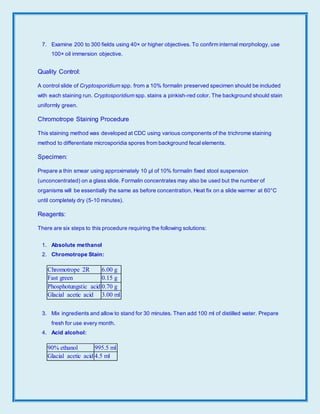
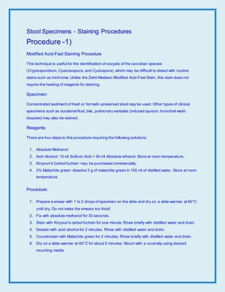
Blood smearThis test is used to look for parasites that are found in the blood. Direct wet mount examination was followed by formalin-acetate concentration Para Pak Meridian Diagnostics Kinyouns modified acid-fast stain. 2 the stability of the stain in solution and on slides.
Stools were routinely investigated for coccidian parasites in the microbiology laboratory in the summer months. The difficulty in controlling Plasmodium vivax the most common cause of human malaria has been complicated by growing drug resistance. The use of larger blood volumes for DNA preparation offers a significant improvement in the limit of sensitivity of detection for the diagnosis of P.
2 Culture-only minority of parasitic infections are diagnosed routinely by culture techniques. 3 the ability to use the stain with carbol-xylol in humid regions. Must dry overnight before staining.
The saline wet mount is an unstained preparation made by using physiological saline. By looking at a blood smear under a microscope parasitic diseases such as filariasis malaria or babesiosis can be diagnosedThis test is done by placing a drop of blood on a microscope slide. The oocysts were mostly diagnosed during a wet mount microscopic examination.
For tissue sections extend time 30 seconds. Rinse in 95 ethanol for 30 seconds. When DNA was purified from 5 millilitres of blood it was possible to routinely detect as few as 50 malaria parasites per millilitre using nested PCR.
Rinse in 90 acid-alcohol for 1 to 3 seconds. A Rapid growth rate - forming a mature. 1 Used in the identification of malaria parasites trypanosomes and microfilariae.
Following assay validation 54 cord blood units donated by mothers who were positive for anti-malaria antibodies were screened by this approach. State the proper method for performing the scotch tape preparation. 5 ml of blood with a parasite density of 50 parasites per ml will contain.
Microscopic examination of Giemsa-stained blood films remains a major form of diagnosis in malaria case management and is a reference standard for research. Take extra care during this step to achieve correct staining of spores. Avian malaria in Brazilian passerine birds.
Falciparum parasites using nested PCR. Any parasite if present can be identified on the smear. Automated image recognition based on.
Parasitism detected by nested PCR using DNA from stained blood smears - Volume 130 Issue 3. Stains or dyes contain salts made up of a positive ion and a negative ion. In a parasitology laboratory routinely two preparations of each specimen are usually made on each slide.
The other uncolored ion is called the counterion. Relatively few of protozoa can be cultured in a manner that is useful for laboratory identification. One unstained preparation and another temporarily stained preparation.
Adjustments can be made as outlined below. Using such methods and following fixation and sample preparation for electron microscopy EM capture of invading merozoites is consistently a rare event making imaging and data collection a. Piroplasms are globally distributed obligate intracellular hemotropic parasites of vertebrates from the genera Babesia Theileria and Cytauxozoon 1 2The name piroplasm comes from the fact that within erythrocytes the parasites often appear as pear-shaped on Romanowsky-stained cytological slides following microscopic examination Microscopic.
Rinse twice 30 seconds each time in 100 ethanol two separate containers are required for this step. 2 Preparation of smear - 3 - 4 drops of blood stirred together to size of a dime. In addition to fixation staining is almost always applied to color certain features of a specimen before examining it under a light microscope.
Check the pH of buffered water routinely before use. The slide is stained under a microscope. Stains and dyes are frequently used in histology microscopic study of biological tissues and in the medical fields of histopathology hematology and cytopathology that focus on the study and diagnoses of disease at a microscopic level.
Polyvinyl alcohol PVA containing the fixative mercuric chloride is considered the gold standard for the fixation of ova and parasites in the preparation of permanently stained smears of stool specimens. Since there are many causes of GI infections an OP may be used in conjunction with other tests such as a gastrointestinal GI pathogens panel or a stool culture. Identify parasites present on a scotch tape preparation.
Examination of a stained peripheral smear is an integral part of laboratory evaluation of patients. We have established a method to cycle parasite generations in continuous culture using human blood cells. Newsprint just legible through smear.
This was especially true of protozoan parasites. It provides information on red cells leucocytes and platelets and is used to supplement the information provided by automated hematology analyzers. Chesson strain parasites were passaged from owl monkey erythrocytes to human reticulocytes in McCoys 5A.
The use of larger blood volumes for DNA preparation offers a significant improvement in the limit of sensitivity of detection for the diagnosis of P. The following is the method for using the Lovibond comparator for. The ova and parasite OP exam is used to detect the presence of parasites in your stool sample and help diagnose an infection of the digestive system gastrointestinal GI tract.
Use the following terms to describe the growth rate.

Pdf The Buffy Coat Method A Tool For Detection Of Blood Parasites Without Staining Procedures

Images Of Relatively Large Blood Parasites Larger Than Red Blood Download Scientific Diagram

Pdf The Buffy Coat Method A Tool For Detection Of Blood Parasites Without Staining Procedures

Main Procedures Of Blood Sample Preparation For The Application Of Download Scientific Diagram
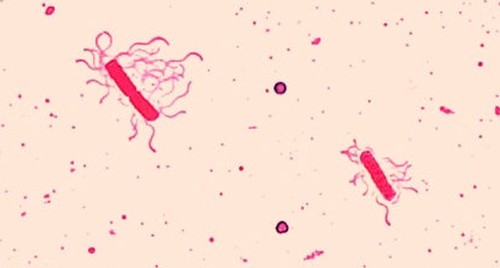
Staining Microscopic Specimens Microbiology
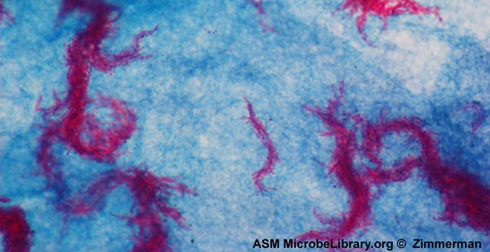
Staining Microscopic Specimens Microbiology

Common Fixatives Used To Preserve Ova And Parasites In Stool Download Table
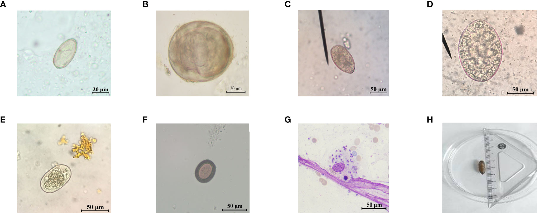
Frontiers Foodborne Parasites Dominate Current Parasitic Infections In Hunan Province China Cellular And Infection Microbiology
In Clinic Hematology The Blood Film Review Today S Veterinary Practice

Pdf The Buffy Coat Method A Tool For Detection Of Blood Parasites Without Staining Procedures

Public Health Risks Associated With Food Borne Parasites 2018 Efsa Journal Wiley Online Library
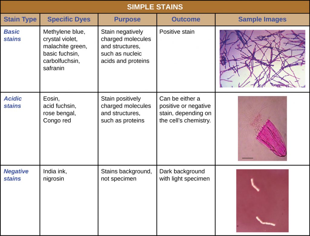
2 4 Staining Microscopic Specimens Microbiology Canadian Edition
Comments
Post a Comment